Heel and foot pain, Plantar Fascia, Part 3. The overlooked tear and strain.

The plantar fascia can be injured in a variety of manners. Most people are familiar with the idea of a repetitive injury involving straining of the fascia at the heel origin, starting a vicious cycle of irritation followed by scar tissue formation, then thickening and scarring of the ligament layers, causing the inflexible tissue to be less and less resistant to walking, and weight bearing activities. The natural instinct in that case becomes to vigorously stretch and manually mobilize those structures (standing calf stretches for example). However, the arch ligament is also subject to partial, acute or sub-acute tearing, making it more akin to a nasty, unstable ankle rolling sprain. The patient will report a rather rapid onset of acute heel pain after a long run, a long day of running errands while wearing flimsy shoes, and the pain is often unilateral. Palpation findings will show a discreet area of thinning of the ligament close to the heel and almost always on the inside of the foot. In extreme cases, you can palpate a gap in the fascia and see a visible small lump under the arch where the torn ligament retracts.

Distinguishing an acute fascial tear and injury is extremely important since many self care approaches used for the chronic phase are contraindicated, especially weight bearing calf stretches. Over the long term you still need to evaluate all of the structures we talked about last week for predisposing the plantar ligament to rupture by pre-loading it excessively, however in the short term you treat it like a partially torn ligament with rest and some degree of immobilization. In routine cases, that will involve compression bandage, limited weight bearing, rigid taping in a high arch position, and a soft arch support. Ultrasound therapy can also be really beneficial for the first couple of weeks after the injury.

In the long run, most of the fascial tears will turn into a chronic case with scar tissue filling the gap, but the integrity of the arch may be compromised due to lengthening of the fascia and weakening of the medial aspect. Formation of a bony spur around the tear is often visible on X-rays within 12-18 months after a tear. Most people will end up needing custom orthotics long term.

Heel and Foot Pain, Plantar Fascia, Part 2.

When a patient presents with heel and arch pain, you need to evaluate several structures that can contribute or cause the problem. This will include examining the following:

  • Structure of the foot arch for integrity or collapse as well as the ankle subtalar joint for pronation. If the joint/ligament complex of the foot or ankle is no longer intact, you will need to address it fairly soon or else the soft tissue injury will not resolve out of the acute stage. This will often require orthotics combined with the right shoe.

  • Posture and alignment of the spine and pelvis. Pelvic rotation or short leg can cause asymetrical stress on one foot. Anterior posture of the neck and upper back can cause overload of the posterior heel ligaments.

  • Muscle balance in the lower leg, especially the deep calf muscle layers. The tendons of those muscles are found on the plantar aspect of the foot and can be over stretched if the calf muscles are unusually tight.

  • The soft tissues of the bottom of the foot, especially the plantar fascia proper. It is absolutely crucial to assess the texture of the plantar soft tissues to determine if the patient is in more of a chronic stage, with thickened, hardened scar tissue formation, or if the patient is in a fresh acute stage with thinning and partial tearing of the fascia close to the heel origin.

Heel and Arch Pain, Plantar Fascitis. No one size fits all approach. Part 1

I have meant to write about this for a really long time but, as the saying goes, it is kinda complicated and I sometime lack the mental energy to write about complicated things.

Heel and foot pain, and “plantar fascitis” are excessively common. And common medical problems are obviously talked about a lot. The down side of that is that they are not always talked about or much less understood correctly. “Plantar fascitis” is a close contender to “sciatica” as a misused term that in reality covers more than one problem. And in the case of plantar fascitis, as in sciatica, knowing more exactly what you are dealing with is pretty important because they may require drastically different treatments.

For starters, you need to understand a bit about foot anatomy. If you peel back the layers from the bottom of your foot (for now, stick with doing that in your head only…), you will encounter a complex layer of ligamentous fascial tissue that has various attachments in the bones of the mid foot and toes, and for the most part, one common origin in the front of the heel bone called the calcaneus. The latter area is where you will have a large portion of your problems and symptoms.

Past the fascial and ligamentous layer, you will encounter several layers of muscular tissues. Some of those muscles start in the posterior calf and only their tendons are present in the sole of the foot; some muscle have their origin and attachments in the foot proper and are called intrinsic muscles.

Past the muscle layer, you will encounter the actual bottom of the bony arch of the foot, comprised of a series of bones arranged in the loose shape of an arch, connected in complex articulations that are designed to “give” under normal weight bearing conditions.

To be continued next week…

More bad news on vaping, e-cigarettes

The headlines of a few days ago was hardly a surprise, but in the midst of an epidemic of youth vaping with the false impression that it is safe, a good reminder that e-cigarettes are turning out to be deadly in their own right. It turns out that inhalant concentrated nicotine in that form is really damaging to the brain, and in much faster acting ways than cigarettes. And younger brains are much more susceptible. Please advocate for the complete ban of e-cigarettes on the same level as regular cigarettes to limit exposure to concentrated inhalant nicotine for everyone.

https://www.nbcnews.com/health/kids-health/fda-warns-about-possible-risk-seizures-associated-e-cigarettes-n990446

vaping.jpg

Inflammation, pain, and extra weight

I have had a couple of touchy discussions with patients last week about addressing all of the issues contributing to their chronic pain and inflammation status. As with most touchy discussions, they revolve around lifestyle issues that patients are often struggling to change or prefer to be in denial about.

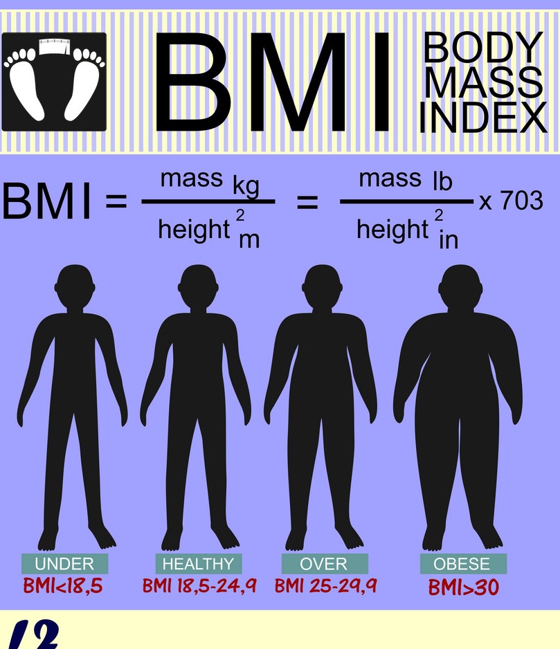
One of those discussions revolved around the role of excess weight. The stats are telling us that our collective BMIs are going up, and I certainly can attest to that in the microcosm of our office. Patient can begrudgingly understand the mechanical impact of excess weight on a downstream joint complex. What is overlooked in the process is the contribution of excess adipose tissue (stored excess fat), in powerfully adding to the pro-inflammatory chemical soup of our bodies. Body fat is far from being an inert blob hanging on to your belly or thighs. In reality, it is a very active endocrine and metabolic tissue that secretes a variety of chemicals. One of those chemicals is called “adipokines”. These substances modulate inflammation control. When body fat is unregulated due to increased storage, the secretion of “pro-inflammatory” substances is elevated and pretty much never goes down until the body fat levels starts to return to a body fat percentage that is more normal. So in individuals with elevated BMIs (AKA overweight or obese), there is an increased likelihood of chronic inflammation affecting joints, ligaments, cartilage, brain, heart, and that compounds the mechanical effect of the excess weight. For those individuals, shifting to a more anti-inflammatory diet pattern, while you slowly work out your weight issues, and possibly adding some anti-inflammatory supplementation to your regimen, may be necessary to mitigate chronic pain.

https://www.ncbi.nlm.nih.gov/pmc/articles/PMC4970637/

body-mass-index-and-silhouettes-of-different-vector-9353473 (2).jpg

Quads and chronic postural low back pain

Quads are a big muscle group that is often below our radar. We use them every day every time we walk even a few steps, but since it does not often cause direct pain, we can overlook the way it can contribute to problems in other areas.

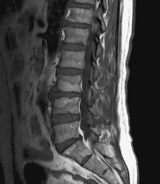
One of these “other areas” would be the lumbar spine. The upper fascial layers of the quads extend into the hip flexors deep in the anterior lumbar spine. The hip flexors and the muscles in particular bear much of the blame for causing anterior lumbar spine shifting and secondary compression/extension of the lower lumbar discs, but the quads can do the same thing. The diagram does a fairly good job at depicting the shift in the lumbar posture. Incidentally, this also causes some overload lengthening of the hamstrings and could be a silent source of constant straining of the posterior thigh.

Heavy use of the quads during certain type of occupations (construction workers, laborers etc…), with little rest in between, seems to be a common trigger for chronic myofascial shortening of the quads (whereas muscle shortening is more often triggered by prolonged sitting). You can pick up on the problem when placing the patient in a stomach lying position and bending both knees to 90’. With normal quads flexibility you should be able to achieve that without causing recruitment of lumbar extension or lateral shifting of the glutes. In patients with short quads, you will start noticing some strong resistance, lumbar extension and side gliding of the thigh as early as 45’.

For patients with very short quads, traditional stretching may not be the place to start since you can further aggravate lumbar extension in the process. You may need to start with more passive lengthening therapies and adhesion break up using a foam roller in the prone position for the entire length of the muscles.

Guillan-Barre Syndrome- Acupuncture and Chiropractic

The people reading this blog are going to fall squarely in two categories: those who will raise an eyebrow and a “huh?”, and those who will really pay attention. I don’t often blog about lesser known medical topics, but after a couple recent patient encounters, thought it would be worthwhile this time.

Guillan-Barre is an acute auto-immune peripheral nerve disease, usually post-viral. It can cause profound, severe and rapid loss of motor function in the limbs, face, spine, as well as profound loss of sensation and tingling. While most patients eventually recover, some continue to have persistent weakness in limb or face, and commonly continued neuropathy in the hands and feet, for years after the acute illness. Beyond the initial medical treatment with immunoglobulins, and rehab, patients do not have a lot of medical options for the residuals.

Acupuncture and chiropractic combined, along with an anti-inflammatory diet and some target supplementation, can make a big difference in the residual symptoms of Guillan-Barre. All of us field practitioners would like to see more published large scale studies, but til then, here is a nice case study

https://www.sciencedirect.com/science/article/pii/S2095754817302272

Guillain barre.jpg

Chiropractic as Part of an Anti-Opioid Strategy

https://www.ncbi.nlm.nih.gov/pubmed/29843912

This great piece of 2018 research has probably not received the publicity it deserves, especially not in light of the sobering stats released from the last 2 years on opioid related deaths in the US, showing little abating. The prescription medications that a patient receives at the time of a first major injury or other musculoskeletal event can be a “gateway drug” to start an addiction cycle that leads patients to street drugs and their lethal potential. And so the need to look at all first line non-opioid interventions in those cases. I rest with a little comfort that our profession is doing their share in trying to reverse the tide.