
Recently I've been looking at a lot of MRIs with patients, trying to go through each line of the report and make sense of the medical jargon. One term that seems to be completely foreign to most patients is the mention of Modic type I and Modic type II changes.
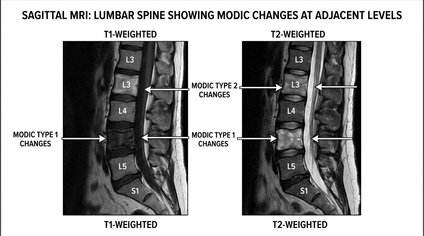
Modic changes refer to some abnormal signal on the bone marrow of the vertebrae adjacent to a spinal disc. They look like abnormal coloring on the top and bottom of the vertebrae. They represent some changes in the normal bone and bone marrow with some infiltration of inflammatory cells, edema. Modic changes are the reflection of the severity and active nature of degenerative changes of the vertebral segment, where the disc degeneration starts to progress to the point of involving the adjacent bone. Modic type I changes are acute, fresh, active, and almost always correlated with active bone pain, whereas Modic type II changes are more of the chronic, potentially non-symptomatic scar tissue of a previous acute episode. The importance of noting those on an MRI is that they tend to be much more correlated with active pain than certain disc changes, especially disc bulges, which can be found at a high prevalence level in the general population but can be completely asymptomatic.
You have to remember that MRI images are extraordinary at giving you a lot of information, including pretty much everything that's ever happened to you but doesn't help you differentiate what's relevant to your particular current complaint. The presence of Modic changes, especially type I Modic changes, can help you differentiate between background degenerative findings versus an active problem. How you treat Modic one changes is more complicated than the intent of this short blog, but does need to get to the root of the mechanical stress to the affected segment and sometimes involve oral supplemental anti-inflammatory control, whether pharmacological or botanical.
