FIBROMYALGIA, CHIROPRACTIC AND THE UPPER CERVICAL SPINE

https://pubmed.ncbi.nlm.nih.gov/25782585/

The topic of fibromyalgia can be daunting because it's broad and complicated. Fibromyalgia remains a diagnosis of exclusion, although over time we’re starting to identify it as a constellation of symptoms that tend to occur together and usually in targeted populations (women in particular.)

Chiropractors routinely see patients with a fibromyalgia diagnosis as part of their practice. That is because fibromyalgia patients can have very discrete neuromusculoskeletal lesions amenable to chiropractic care that if left unattended, can greatly amplified the chronic background pain.

This article explores another aspect in which chiropractic care can fit in the overall treatment plan of patients with fibromyalgia, and this time from a very different angle. Research in fibromyalgia of the last 10 years has unearthed multiple mechanisms of causation, including some degree of autonomic nervous system dysfunction. The upper cervical spine is extremely rich in sensory afferents to the brainstem, where most of our autonomic control is located. Spinal functional lesions in the upper cervical spine does have a greater impact on autonomic dysregulation, and potentially a great therapeutic impact with chiropractic correction in fibromyalgia patients.

One item of note: fibromyalgia are very slowly changing conditions over time so treatment plans need to adapt to that reality and involve slightly more spread out treatment over a longer period of time to be effective.,

fibromyalgia.jpg

Pregnancy Support & Tape

While looking for a less bulky and more consistent support during pregnancy we decided to give taping support a try. I have been familiar with taping different injuries for tissue offload, support or proprioceptive changes, but I haven't had a lot of chances to work on taping during pregnancy. In the images you can see a tan tape wrapped around the low abdomen pulling up and back to assist in the pulling forward mechanism that a growing baby imposes on the low backs of many pregnant women. What you may not be able to tell is this, but this particular tape application also is having more tension on the right and we were able to place the tape in a particular stretch position in order to help offload and decrease that tension on the right side and distribute towards the left. 

Some pregnant women may be familiar with the braces, bands or scarf techniques to help offload, but if you've used many of them you’ll notice they tend to be bulky & uncomfortable. Sometime you may even need to redo or adjust throughout the day. Having a tape placed directly to the skin that tends to last a number of days may be a better option. You can even shower with the tape on!

OSTEOPOROSIS PART 6

On the final segment of the osteoporosis series, I wanted to answer a question that often comes up in relationship to osteoporosis and briefly talk about nonpharmacological approaches.

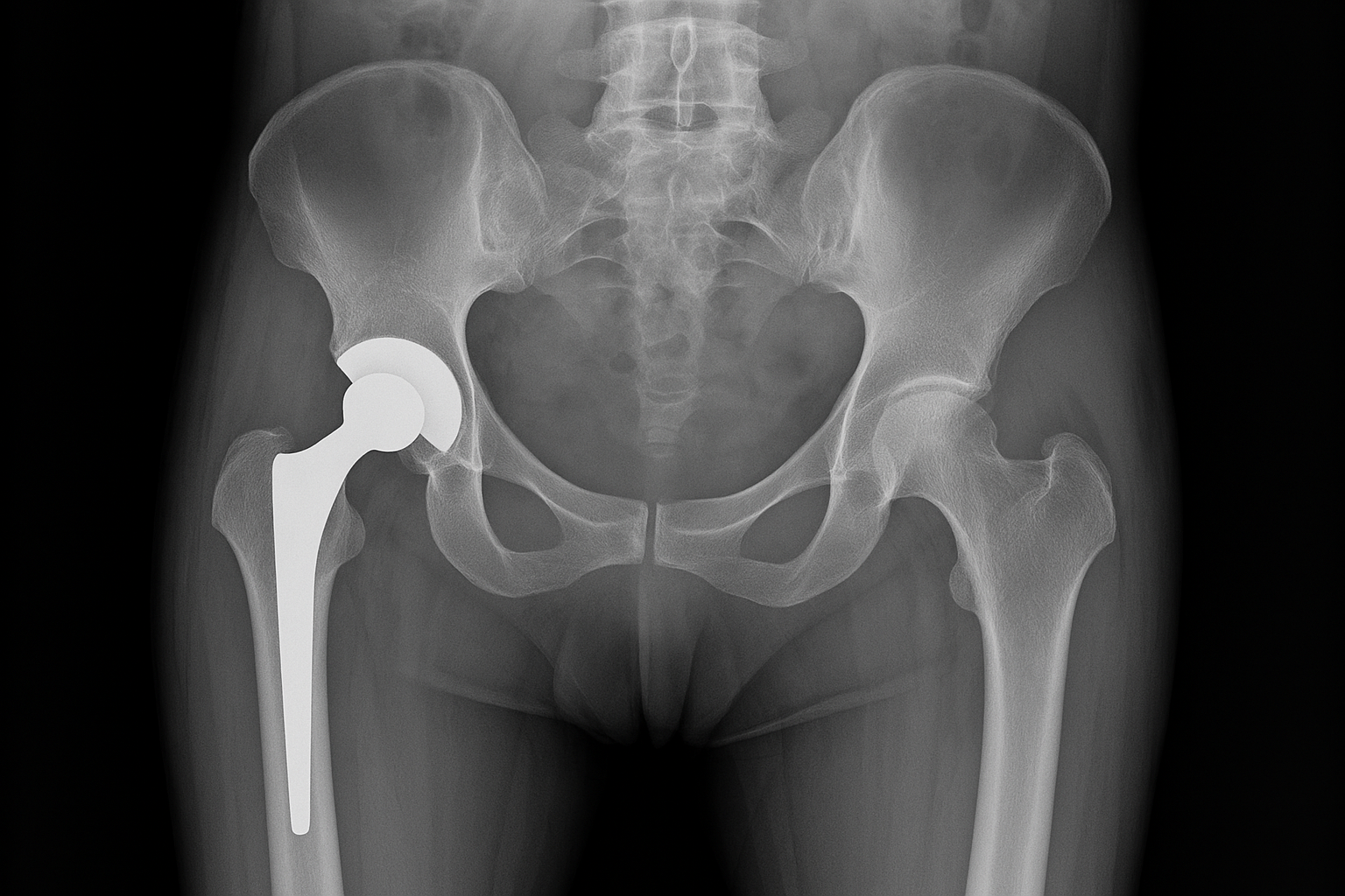
Patients often ask if osteoporosis by itself can be pain producing. I think that in most cases the answer is no and that is the general traditional medical understanding. Obviously complications of osteoporosis like stress fractures can be extremely pain producing, but as to the question of how symptomatic a low bone mass can be, the general understanding has been that it is silent. However I would say that in the last five years I have started shifting my thinking on the subject after encountering several patients with fairly advanced osteoporosis with T-scores well below -3.5, who reported some generalized vague bone pain, not related to position or movement, and I have started to wonder if in some extreme cases, osteoporosis can give you some non- localized symptoms. In most cases however, when a patient presents with back pain and osteoporosis, the source of the pain is going to be mechanical, postural, myofascial, degenerative or inflammatory in nature.

As far as treatment options, the recommendations are obviously going to be based on the results of the workup that we have described in the first five segments.

We would first address some of the basic bony needs such as adequate dietary intake of bone matrix nutrients (protein, calcium, minerals,) as well as adequate weight-bearing resistance training to stimulate the body's deposition of bone.

Second, we would try to address and correct some of the abnormalities found on lab testing: treat digestive inefficiencies such as hypochlorhydria, significant menopausal hormone deficit, elevated chronic stress hormones, etc. The limitation is sometimes the medications that the patient have to continue taking long terms. In which case it may not be fully possible to correct the underlying problem, and more aggressive supportive supplementation is needed.

Third, nutritional supplementation and intervention has to be catered to a particular patient's need. There is not a one-size-fits-all supplementation for all patient with osteoporosis/osteopenia. But in most cases, the patients will need to make sure they have adequate intake of vitamin D3 and vitamin K2. The exact dosage varies but usually ranges somewhere between 2000 and 5000 IUs daily to achieve a healthy blood level around 30/40. Vitamin K 2 can be used in physiological doses, similar to what would be a normal dietary intake and absorption around 800 µg, but can be used in more aggressive therapeutic doses of a few grams a day, especially in patients who are exhibiting significant elevation of D-pyrilink on urinary excretion testing.

Cofactors and minerals are extremely important for the body's ability to properly incorporate calcium into the bone matrix, so most commercial supplementation will often contain additional things such as magnesium, boron, strontium. It is also important to note that not all forms of calcium are created equal for the sake of absorption, and that higher-quality supplementation will typically contain bioavailable forms of calcium rather than calcium carbonate. And since calcium is notoriously hard to digest, especially with age, due to decreased stomach acid production, taking the calcium with mildly acidic food can be very beneficial to improve gastrointestinal uptake.

Taking a look into the tool box for soft tissue care

In past blogs we have discussed different techniques and uses for soft tissue care and why it is important. I was thinking having an idea of what each of these instruments actually look like may be helpful as well.

Tennis ball- mostly used for at home soft tissue care.

Tape- there is one whole blog on this, but to recap. In this office kinesiology tape & biomechanical tape is most frequently used.

Resistance bands- used for at home care and activities for continued development.

IASTM & Cupping- there is also a blog published on this awhile ago. This is used mostly in the office and the different instruments I use include a few different stainless steel options that I use for Instrument Assisted Soft Tissue Mobilization (IASTM) as well as either some silicone or plastic cups for cupping. The IASTM provides more of a compression as where the cupping creates more of a pulling or suction to the targeted underlying soft tissue.

What are some of your favorites?

SOFT_TISSUE_TOOL_BOX.png

OSTEOPOROSIS PART 5: IT IS COMPLICATED, HOW YOU GET IT, WHAT YOU DO ABOUT IT

Today I would like to expand on the last discussion and discuss a more comprehensive testing strategy beyond DEXA scan to look for root causes of osteoporosis.

The first additional testing that I would consider when someone comes in with questions about recent DEXA score would be a D-pyrilink urinary test. The D-pyrilink is a protein that is part of the bone matrix. During the process of bone break down and recapture for new bone formation, the D-pyrilink protein is briefly circulating in free-form and can be excreted through the urine, and the amount can be measured. If the bone is reabsorbing and being rebuilt at a normal rate without excessive loss, the amount of D-pyrilink in the urine will be relatively modest. If the body is breaking down bone faster than recapturing and rebuilding, the amount will be elevated. So measuring urinary D-pyrilink excretion can be a useful addition to a DEXA scan to see if the active state of bone loss.

Additional tests that can be helpful in circling in on the causes of a person's osteoporosis and bone loss includes:

– A hormone panel to assess the adequacy of estrogen, progesterone, testosterone, cortisol, and DHEA (basic anabolic adrenal marker). Incidentally most of the commercially available D-Pyrilink tests are lumped together with the above hormones in a single bone health panel.

– A vitamin D metabolite panel. This would include not only the commonly tested D3, but a metabolite called calcitriol, which is directly converted from vitamin D3 in response to the body's needs to improve calcium absorption in the gastrointestinal tract. This can be a useful indicator of the body’s perceived lack of available calcium, either from inadequate dietary intake or difficult absorption for a variety of reasons. In which case the ratio of calcitriol to vitamin D3 will be elevated. In addition, we will often get a basic parathyroid hormone level. The parathyroid hormone is the master control of calcium blood level regulation. Under normal circumstances with adequate vitamin D/calcium economy, the parathyroid levels will be in the lower end of the range. Even slight elevations of parathyroid hormone still considered normal but suboptimal can indicate the body's perception of inadequate calcium/vitamin D economy.

– A gastrointestinal functional markers profile. This can be very useful when we suspect that the patient has adequate dietary intake of nutrients necessary for bone formation such as protein and calcium, however the body is unable to properly absorb those nutrients for variety of reasons (prior abdominal surgeries, chronic infections, hypochlorhydria, mild digestion, inflammatory bowel disease etc.) the panel can measure stool residues of various digestive enzymes and nutrients, telling us if absorption is the problem causing osteoporosis.

– Last but not least, and probably first and foremost in most cases, a good diet history to see if the patient has a reasonable amount of nutrients available for normal bone formation.

TENNIS RELATED INJURIES & HOW YOUR DAILY WORK MAY BE SIMILAR

While reading up on some upper extremity injuries associated with sport, I came across this piece from Epidemiology of musculoskeletal injury in the tennis player. Based on information found the authors are hypothesizing that “teaching the topspin serve at a young age may put the young tennis player at increased risk for back pain and/or injury.” It is thought that “early introduction to the topspin serve may be associated with the development of spondylolysis and/or spondylothesis in elite-level players.”

This particular piece of research goes into other injuries associated with tennis such as lower extremity pains, association with volume of play, age and sex, skill level and even racquet grip position. 

Racquet grip position is another one I found to be very interesting as different grips have been associated with different overuse injuries.

  • Ulnar sided injuries (extensor carpi ulnaris tendonitis and triangular fibrocartilage complex pathology) were significantly associated with a Western or Semi-western grip

  • Radial sided injuries (flexor carpi radialis tendonitis, De Quervain's tendinopathy and intersection syndrome) were more common with Eastern grip.

tennis-grips.png

With this information regarding tennis racquet holds being associated with different overuse injuries, we can see how this may relate to daily work , job dependent. Below you can see the photo of how this individual holds a rod, what you don’t see is that they have a large GPS unit above that needs to be stabilized, similar to stabilization needed when hitting a tennis ball. Some different wrist and hand positioning on this rod may pre- dispose this individual to these same overuse injuries.

IMG-6403.JPG

Which tennis grip style is most like this positioning?

Osteoporosis Part 4: It's Complicated. How You Get It, What You Do About It…


In this next part of the ongoing discussion regarding osteoporosis I wanted to talk about diagnosis and the confusion that surrounds it.

Standard medical diagnosis of osteoporosis or osteopenia is done by a low-dose radiation x-ray of the hip and spine call a DEXA scan. Please note that plain film x-rays are not a good way to screen or establish a firm diagnosis of osteoporosis although it will often indicate some issues with bone mass that need to be followed up with the appropriate diagnostics.

The results of a DEXA scan are reported as the T score and Z score in both the hip joint and in the lower lumbar spine. A mild decrease in bone mass compared to what is considered standard will result in the diagnosis of osteopenia while a more significant decrease of bone mass will result in a diagnosis of osteoporosis. The scoring associated with a diagnosis of osteoporosis is also usually associated statistically with a certain fracture risk over the following 10 years.

As a patient you need to understand that there are some caveats and limitations associated with how you interpret your own DEXA scan results.

- The DEXA scan has a certain margin of error. If you are close to a cutoff of osteoporosis or osteopenia this could be significant.

- The margin of error can be amplified when comparing repeat scans by having the procedure performed on a different machine or by a different technician. In some instances this could be significant and make the interpretation of comparative scans difficult. If you're tracking osteoporosis response to treatment whether medical or integrative, you really should always be getting your scan on the same machine at the same facility.

- There are some issues with the spine and hips that can give you a false positive or negative readings, most commonly positive readings. This is especially true for people who have some degree of degenerative disc disease with bone spurs, more advanced degeneration, or congenital anomalies of the lumbosacral spine such as a spondylolisthesis. If this is a significant concern for false-positive, a quantitative CAT scan would be the definite testing but it involves more radiation.

- There is some question about the validity of a single scoring system applied to different ethnic groups, as well as in women with very small frames, whose bone mass may be naturally and sufficiently lower than their counterparts. It is also true that women with very high BMI may look like they have adequate bone mass but in reality the bone density may not be sufficient to protect them from fracture because of the increased demands associated with the extra weight.

- The DEXA scan measures the raw amount of bone mineral, however it does not measure the quality of the bone architecture which is very important for fracture prevention. This is not to invalidate the benefit of a DEXA scan but to realize that there is still some question about whether or not current osteoporosis drugs improve bone density without concurrently improving architecture, meaning that we may get a false sense of security against fracture risk long-term.

- Finally and possibly most importantly the DEXA scan only measures one snapshot of your bone mass in time. Unless you have comparative scans you have no idea if you are currently losing bone, are in the process of starting to lose bone, or have previously lost bone mass but have plateaued. This is why I strongly suggest that every woman has a baseline DEXA scan prior to menopause or at the onset of menopause for future comparison and good treatment decision.

- Based on the above, it can be extremely useful to couple the DEXA scan with other testing that may give you more real-time information about bone metabolism and turnover. One such test which is mostly used in integrative health practices rather than allopathic traditional medical care would be a urinary D-Pyrilink excretion test. We will talk more about it in our next segment.

165246418_2819833671573214_2591375601920367480_o.jpg


Taping

Recently I’ve been getting more questions regarding taping, different techniques and the differences between tapes. The main tapes I have become familiar with are white tape or athletic taping or strapping. This is the tape that most people are familiar with on ankles in football. This tape is mostly used for support and structure. I don’t tend to use this tape very often in the clinic. Kinesiology tape is the next most common tape people know. This tape tends to be seen on athletes everywhere and has been around for awhile. I tend to use kinesiology tape mainly for some short term movement facilitation. Often patients will note feeling some additional support. The last tape I’m going to write about is a “newer” type of tape, dynamic tape. This tape is reported to be more of a biomechanical tape. I use this a lot in clinic for offloading techniques. Meaning when an injury occurs many times some of the load or weight needs to be taken off the injured tissue in order to allow for optimal healing. There are ways we can use this tape to assist in providing some of that offload as well as other in office techniques to assist in the recovery.

tape.jpg