Getting your Pregnant Body ready for Birthing

It has been a while since I have written about chiropractic and pregnancy, because I have been too busy talking about feet, but a crop of pregnant bellies have made their way to the office again, reminding me of this very important topic.

I had a chance to attend an excellent training seminar last month, taught by a very seasoned midwife who has a long history of working side by side with body works practitioners, including chiropractors. As she progressed through her professional career, she started to realize the profound correlation between the neuro-muscular structures of the spine and pelvis, optimal fetal positioning, and successful labor progression. While I have studied the chiropractic approach to pregnancy and have worked on pregnant women for 25 years, I never cease to be surprised by how much more I can learn. Gail Tully’s seminar gave me a more refined understanding of how certain spinal structures determine the progression of various phases of labor and gave me a few new preventive tools as well.

I would highly recommend her website as a resource for pregnancy preparedness. The information is generic and you have to figure out what may apply to your particular situation or not (and working with your local chiropractor can help you devise an individual plan from that material). Here is a link to some daily activities of preparedness that will be safe in most instances.

https://spinningbabies.com/start/in-pregnancy/daily-activities/

Pregnancy Blog.jpg

Heel and arch pain, plantar fascia part 5. Treatment

Heel and arch pain, plantar fascia part 5. Treatment

The treatment of heel, arch and foot pain starts by understanding what structures are involved, how acute or chronic the problem is, and what some of the root causes may be.

If a patient present with an acute ligamentous injury, or an acute flare of a chronic problem, you will need to start by resting the arch and avoiding additional straining: semi-rigid taping, soft standard insoles, cold application, oral botanical anti-inflammatory supplementation, supportive shoes and minimal weightbearing stress. You can start addressing root causes by adjusting the spine, correcting faulty pelvic postures. Stretching of the posterior calf fascia needs to be done actively and non-weightbearing. Ultrasound can be helpful when dealing with a lot of swelling and inflammation ,especially with an inflamed, chronic heel spur.

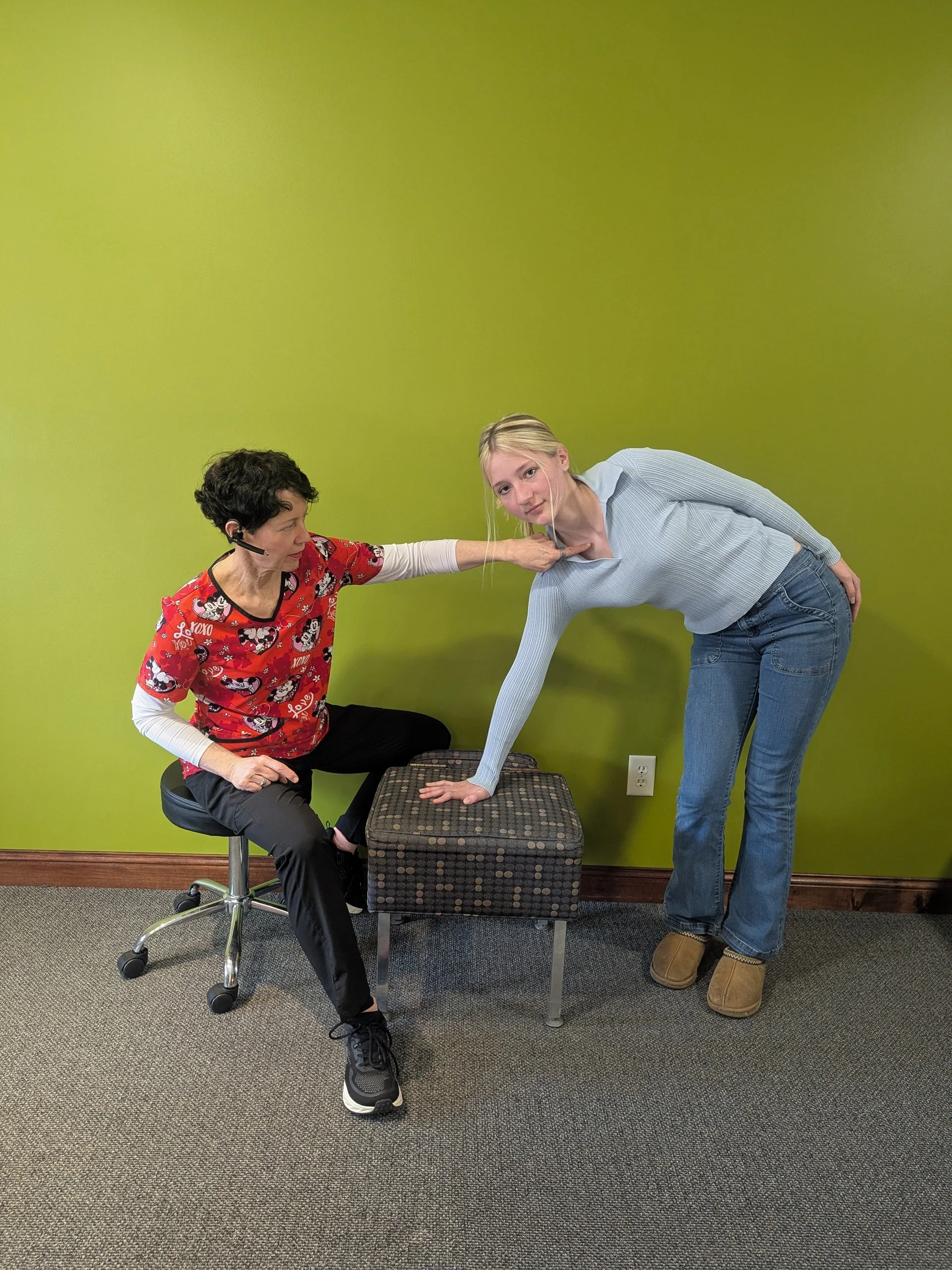
Chronic fibrous plantar fascial dysfunction can tolerate, and will benefit from a more aggressive approach using deep tissue mobilization in various parts of the arch and calf, as well as more intense, weightbearing stretches. Custom orthotics can be introduced once soft tissue flexibility has been reasonable restored, and significant spine and pelvis imbalances have been corrected. Supplementation that improves peripheral blood flow, both topical and oral, are sometime needed. Scare tissue has poor blood flow, and the body cannot easily soften and replace it with more elastic collagen unless there is adequate oxygen concentration in peripheral tissues.

Recovery time can vary, but is almost always slower than a patient would prefer. It is important to remember that most of the pain generating tissues are comprised of ligaments, tendons and fascia, which are less vascular than muscles and have a slower turnover rate. In general you should bank on 6-8 weeks for a first time problem, 2-4 months for chronic cases

WELCOME OUR NEW CHIROPRACTIC SENIOR INTERN KAILA ALVAREZ!

kaila (2).jpg

Big News-

Join us in welcoming our Senior Chiropractic Intern Kaila Alvarez.

Kaila will be working with Dr. Demel over the next 12 weeks.  She will be assisting in the care of Dr. Demel’s patients in addition to working independently in running a Community Sports Care Program and Pro Bono Care Program. We’re excited to have her join us and we hope you will be too! Be sure to share the good news with your friends and family. Click the links below for an application.

Kaila Alvarez is in her final months of Doctor of Chiropractic training at Northwestern Health Sciences University in Bloomington, Minnesota. She grew up in a small town near Madison, Wisconsin with a family that could always be found at a sporting complex on any weekend. After a couple of years living in Bloomington, Kaila and her husband recently moved to Nerstrand. Her goal is to contribute to a community of individuals to reach their peak performance through chiropractic care and quality communication with a medical team, because people deserve to perform at their best. In her previous internships she has had the privilege of providing care for all ages, from high school students to professional athletes. She looks forward to assisting you in achieving your maximum potential!"

Fiona and Kaila (2).jpg

Heel and foot pain, part 4. The heel spur.

Heel spurs are the reason for much confusion. As in the infamous mention of a“disc bulge”, the mention of a heel spur brings on a lot of unwarranted cold sweat.

Heel spurs are quite commonly found on X-rays of patients who are being evaluated for another foot trauma and have no current symptom in the arch of heel proper (I lost track of how many times I see a spur on a film taken after a bad ankle sprain). The reason is that the spur, also known as a calcanear osteophyte in medical term, is the end result of a cycle of repetitive strain and tearing of the plantar fascial common ligamentous origin at the heel. When the repetitive tearing involves micro-bleeding, or a lot of persistent inflammation, the body will deposit calcium over the injured area, resulting in a spur.

The heel spur proper is an indication of a prior injury to that area that has resulted in calcification of the injured soft tissue, since the calcification process takes several months to build up, then show on X-rays. However, while many patients will have a non-symptomatic heel spur, many patients will have continued symptoms over the area. The reason is that in many patients, if the mechanical factors causing the strain over the plantar ligamentous origin at the heel are not addressed, the body will go through a cycle of ongoing re-injury of the area, with the heel spur growing over time.

The examination process in patients with heel pain with or without heel spurs is the same, but the presence of the spur is a definite indication that the problems has been going on for a very long time.

Heel and foot pain, Plantar Fascia, Part 3. The overlooked tear and strain.

The plantar fascia can be injured in a variety of manners. Most people are familiar with the idea of a repetitive injury involving straining of the fascia at the heel origin, starting a vicious cycle of irritation followed by scar tissue formation, then thickening and scarring of the ligament layers, causing the inflexible tissue to be less and less resistant to walking, and weight bearing activities. The natural instinct in that case becomes to vigorously stretch and manually mobilize those structures (standing calf stretches for example). However, the arch ligament is also subject to partial, acute or sub-acute tearing, making it more akin to a nasty, unstable ankle rolling sprain. The patient will report a rather rapid onset of acute heel pain after a long run, a long day of running errands while wearing flimsy shoes, and the pain is often unilateral. Palpation findings will show a discreet area of thinning of the ligament close to the heel and almost always on the inside of the foot. In extreme cases, you can palpate a gap in the fascia and see a visible small lump under the arch where the torn ligament retracts.

Distinguishing an acute fascial tear and injury is extremely important since many self care approaches used for the chronic phase are contraindicated, especially weight bearing calf stretches. Over the long term you still need to evaluate all of the structures we talked about last week for predisposing the plantar ligament to rupture by pre-loading it excessively, however in the short term you treat it like a partially torn ligament with rest and some degree of immobilization. In routine cases, that will involve compression bandage, limited weight bearing, rigid taping in a high arch position, and a soft arch support. Ultrasound therapy can also be really beneficial for the first couple of weeks after the injury.

In the long run, most of the fascial tears will turn into a chronic case with scar tissue filling the gap, but the integrity of the arch may be compromised due to lengthening of the fascia and weakening of the medial aspect. Formation of a bony spur around the tear is often visible on X-rays within 12-18 months after a tear. Most people will end up needing custom orthotics long term.

Heel and Foot Pain, Plantar Fascia, Part 2.

When a patient presents with heel and arch pain, you need to evaluate several structures that can contribute or cause the problem. This will include examining the following:

  • Structure of the foot arch for integrity or collapse as well as the ankle subtalar joint for pronation. If the joint/ligament complex of the foot or ankle is no longer intact, you will need to address it fairly soon or else the soft tissue injury will not resolve out of the acute stage. This will often require orthotics combined with the right shoe.

  • Posture and alignment of the spine and pelvis. Pelvic rotation or short leg can cause asymetrical stress on one foot. Anterior posture of the neck and upper back can cause overload of the posterior heel ligaments.

  • Muscle balance in the lower leg, especially the deep calf muscle layers. The tendons of those muscles are found on the plantar aspect of the foot and can be over stretched if the calf muscles are unusually tight.

  • The soft tissues of the bottom of the foot, especially the plantar fascia proper. It is absolutely crucial to assess the texture of the plantar soft tissues to determine if the patient is in more of a chronic stage, with thickened, hardened scar tissue formation, or if the patient is in a fresh acute stage with thinning and partial tearing of the fascia close to the heel origin.

Heel and Arch Pain, Plantar Fascitis. No one size fits all approach. Part 1

I have meant to write about this for a really long time but, as the saying goes, it is kinda complicated and I sometime lack the mental energy to write about complicated things.

Heel and foot pain, and “plantar fascitis” are excessively common. And common medical problems are obviously talked about a lot. The down side of that is that they are not always talked about or much less understood correctly. “Plantar fascitis” is a close contender to “sciatica” as a misused term that in reality covers more than one problem. And in the case of plantar fascitis, as in sciatica, knowing more exactly what you are dealing with is pretty important because they may require drastically different treatments.

For starters, you need to understand a bit about foot anatomy. If you peel back the layers from the bottom of your foot (for now, stick with doing that in your head only…), you will encounter a complex layer of ligamentous fascial tissue that has various attachments in the bones of the mid foot and toes, and for the most part, one common origin in the front of the heel bone called the calcaneus. The latter area is where you will have a large portion of your problems and symptoms.

Past the fascial and ligamentous layer, you will encounter several layers of muscular tissues. Some of those muscles start in the posterior calf and only their tendons are present in the sole of the foot; some muscle have their origin and attachments in the foot proper and are called intrinsic muscles.

Past the muscle layer, you will encounter the actual bottom of the bony arch of the foot, comprised of a series of bones arranged in the loose shape of an arch, connected in complex articulations that are designed to “give” under normal weight bearing conditions.

To be continued next week…

More bad news on vaping, e-cigarettes

The headlines of a few days ago was hardly a surprise, but in the midst of an epidemic of youth vaping with the false impression that it is safe, a good reminder that e-cigarettes are turning out to be deadly in their own right. It turns out that inhalant concentrated nicotine in that form is really damaging to the brain, and in much faster acting ways than cigarettes. And younger brains are much more susceptible. Please advocate for the complete ban of e-cigarettes on the same level as regular cigarettes to limit exposure to concentrated inhalant nicotine for everyone.

https://www.nbcnews.com/health/kids-health/fda-warns-about-possible-risk-seizures-associated-e-cigarettes-n990446

vaping.jpg