Spinal functional lesions and the risk for disc degeneration

https://chiro.org/Low_Back_Pain/Uneven_Intervertebral_Motion.shtml

I have been taking a long nerd safari recently in a newly discovered chiropractic research website and finding some real gems. This particular research paper caught my eye because it provides some clues to one of many facets of risk for a spinal disc to degenerate. Most patients think of trauma, overuse and age as risk factors. But mechanical inefficiency of a spinal segment ( AKA joint is really “stuck” ) can be a big trigger for single disc degeneration, because the lack of normal motion in a segment deprives the joint of the normal lubrication it needs to repair itself from normal wear and tear. The article was also enlightening in that it describes a method for documenting and quantifying the mechanical inefficiency (different from the more routine assessment methods during patient encounter, mostly based on palpation and plain film imaging), which would not be necessary for clinical care but is necessary for research purposes

LYMPHEDEMA: Common Questions

Lymphedema: Common Questions


This is the first of a group of blogs on lymphedema that I want to write. In later blogs I will probably focus on specific aspects of the disease, treatment, management, etc., so I decided to start with a more general Q and A as a basis for future articles. In a chiropractic and massage therapy clinical setting, most patients that come in with swelling complaints have post-injury or post-surgical swelling that has become chronic. There is a range of treatment with these patients, from conservative to more intensive depending on how their swelling has presented and progressed. This will be discussed in more depth in a future article. The information below is for readers to get a general grasp on what is going on when swelling occurs.

lossy-page1-640px-Circulatory_system_with_labels.tif.jpg


What is lymphedema? Lymphedema is a certain type of swelling that results from dysfunction of, or damage to, the lymphatic system. Our bodies drain water, waste products, proteins, and other substances from our tissues via a network of vessels that make up the lymphatic system. This system is especially designed to pick up, filter, and transport lymph fluid back the heart to reenter blood circulation. If damage occurs to lymphatic vessels or lymph nodes (the immune powerhouses that kill harmful cells, bad bacteria, and viruses found in the lymph fluid) swelling can occur in nearby tissues as a result. Lymphedema is a progressive disease because once a certain level of damage takes place, lymphatic tissue repair can no longer occur.

640px-Blausen_0623_LymphaticSystem_Female.png


Is lymphedema treatable? Yes, it is treatable. It is important to state that lymphedema is not curable; that being said, it has been successfully managed by many patients. The goal of treatment is to get the area of swelling back to as close-to-normal size as possible. This is achieved with a treatment called Complete Decongestive Therapy.


Which people have a risk of developing lymphedema? The people at highest risk for developing lymphedema are those that have had lymph node removal, radiation therapy, or severe injuries. People who have had mild to moderate injuries have a lesser chance of developing long-term swelling, although it is somewhat common. Some people who have issues with poor venous blood return in their legs also may be at risk, because blood pooling causes a higher load of water for the lymphatic system to remove. Over time this can cause the lymphatic system in the legs to become diseased, and swelling can occur.

640px-Lower_Limb_Lymphedema.png


What types of swelling are not lymphedema? Some systemic diseases such as congestive heart failure, kidney failure, and abnormally low blood protein levels can cause water accumulation in the tissues (swelling). In these cases, swelling will always manifest on both sides of the body equally, unlike lymphedema. This is because the lymphatic system is functioning normally, while the underlying conditions are the cause of the swelling.

Images used courtesy of Wikimedia Commons

MODALITIES: PART II- CONTRAST THERAPY

Many times we are asked “should I ice or heat” I personally don’t usually have too much of a preference and it’s usually what seems to be feeling better. In general it’s usually noted that acute inflammation is treated with ice since putting more heat on it could increase the inflammation. I also have the opinion that at the same time there is a reason our bodies are made to have inflammation with an injury, in order to flush out the damaged cells and bring in the good cells for healing. 

What I really like is contrast therapy. Contrast therapy is most easily used at home as baths or ice/heat packs. There are some really fancy pieces of equipment out there that will switch from ice to heat quickly, but with that comes a massive price tag. The contrast baths can be used as two small tubs, one with hot water (100 to 111 degrees) and cold water (50-64 degrees). There is a protocol that reports four to one heat to cold is necessary.

This works as a pumping mechanism. The cold is going to vasoconstrict the vessels (make smaller), while the heat will vasodilate the vessels (make bigger). This is going to create the pumping mechanism of dilation, constriction, dilation, constriction, pumping nutrients, blood, oxygen all the good stuff to the injured area while taking away those damaged cells.


Disc Bulge, Herniation, Prolapse, Tear: What is the Difference ?

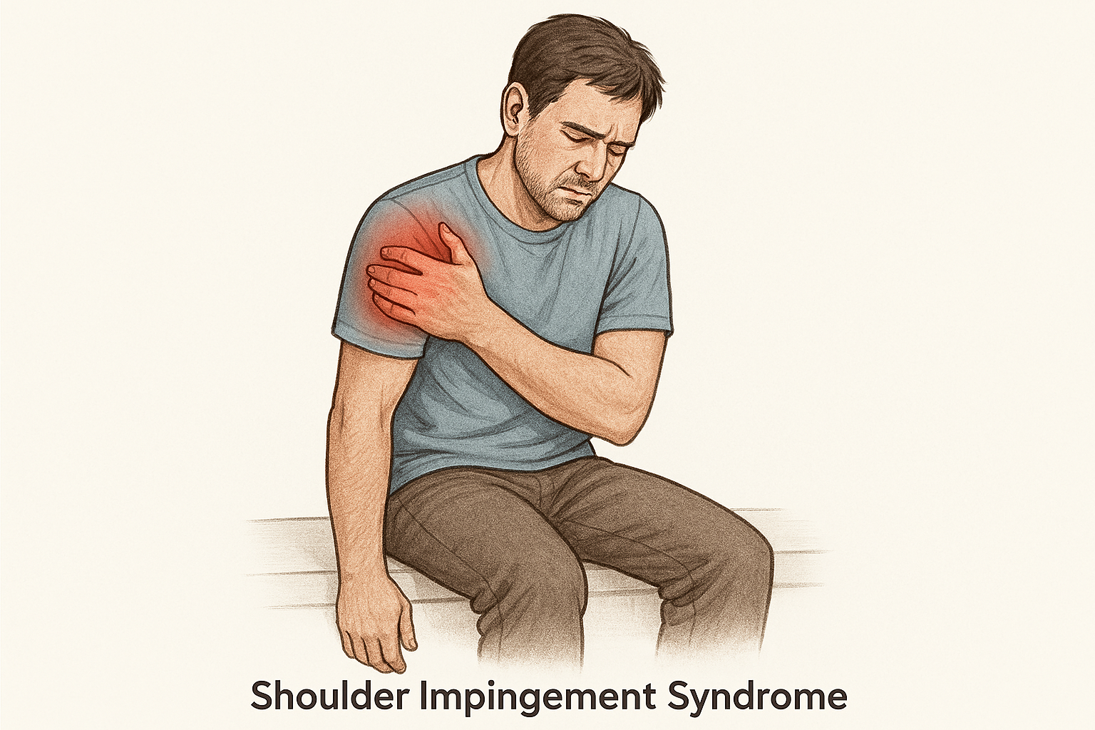
On the long list of items to write about was a clarification of disc pathologies. The term “disc” alone creates sweaty palms in many patients since few understand the spectrum of spinal disc disorders and their clinical significance (and at times lack thereof). It has been relegated to the bottom of the list until now since the topic will have to spread over several blog entries.

First the anatomy: the disc is really an amazing design that allows us to live as bipeds in gravity and with flexibility. It is called a “hydrostatic system”, which means there is an interior gel like component, (the disc nucleus) that is highly hydrated, as well as an outside scaffold of interwoven ligament layers crisscrossing for strength and resiliency to injury. The inside gel resists gravity forces because it is contained in its strong ligamentous wall; at the same time , it can change shape enough during movement to give each spinal segment a certain range of motion, which when combined together, gives you the global range of a spinal region.

An important detail of the anatomy of the disc is the fact that it is bordered by the body of the vertebra above and below, and some disc injury happen at that site (Schmorl’s nodes when the nucleus gel “cracks” into the body of the vertebra during high top down forces). Equally important is the fact that the outside ligamentous shell attaches very strongly to the outside rim of the vertebra above and below. This disc as a whole cannot “slip”: the “slip” terminology is a lay term and imagery, but the anatomical reality involves the disc nucleus gel to breach through and protrude through the ligamentous shells to various extent and degrees of severity.

Another important detail of disc anatomy is the fact that the outside ligamentous scaffold has areas of relative vulnerability at a 45 degree angle posterior lateral, where the shell can be more easily breached, leading to some displacement of the gel nucleus and possible bulging or herniation. Incidentally that is also the anatomical area where the disc is in closest proximity with the exiting spinal nerve of that segment, and the reason that a disc herniation can cause pain to radiate into the leg, along the path of the compressed nerved. The area of vulnerability is more exposed to breach when the spine is loaded with a weight, in flexion (exposing the posterior shell to increased stress), and when the spine is rotated ( the diagonal crisscrossing of the ligaments is less resistant to tear during rotation). That the classic description of an acute disc injury when someone is bent forward, twisting and lifting at the same time.

The types of injuries and pathologies of the disc are quite varied. While in laymen’s term a patient will often describe “something wrong with a disc”, health care providers need to be more specific to understand what exactly is happening to a disc.

How do disc problems develop?

TO simplify things I would say that there are two main mechanisms by which you can develop a disc pathology:

  • Acute disc injury: the typical lift/bend/twist injury, the fall, the hit etc… These injuries definitely occur and patients are very clear on the onset of the problem as a defined event. Remember that the discs are attached to the vertebra above and below via the margin of the ligamentous shell, so the disc injury always happens in the context of a traumatic injury to the entire complex of the two vertebrae framing the disc in question, which is why chiropractic intervention can be so effective in acute disc injuries by addressing the proper alignment of one vertebra in relationship to the other

  • Chronic mechanical abnormal load and stress on the disc, leading to repetitive straining of the disc ligament over time. This is probably the more common cause of disc injury. Often patient report a sudden onset of acute pain, but when you question them further, they will actually describe a rather chronic situation of constant discomfort, stiffness in the morning, soreness after work, not severe enough to seek care but still noticeable. The sudden onset of acute pain is the proverbial “last drop in the bucket” of a progressive problem. Chronic mechanical stress loading of the disc is secondary to other issues: chronic poor posture and abnormal spinal alignment, repetitive strain activities, abnormal core strength, poor muscular balance and flexibility.

What then happens to the disc structure?

In order to answer that question it is helpful to understand certain terms that describe disc pathologies.

A disc bulge: the posterior ligamentous shell of the disc is bowing out, protruding beyond its normal margin. Bulges can be acute and traumatic, when the trauma causes the gel to partially stretch or tear the inside of the shell but not to the extend of breaching through. However, the MAJORITY OF DISC BULGING IS NOT TRAUMATIC, BUT AN INDICATION OF GRADUAL DETERIORATION OF THE DISC OVER TIME. This is an area of great confusion for patients. Besides the patient history, plain films can give you a clue by showing some relative loss of disc height, and MRI imaging can more definitely confirm the source of a bulge: the slowing deteriorating inside gel “dehydrates” ( basically dries up), causing the two vertebrae to approximate and the ligamentous shell to bow out.

herniation-v-bulge-v-normal-back-disc.jpg

A disc HERNIATION: the inside gel breaches through the posterior shell. That phenomenon alone can be quite painful locally. Depending upon the exact location of the disc gel fragment, it may or may not come in contact with a neurological tissue such as a spinal nerve going down the leg. Herniations are more often the result of an injury, although they can become chronic or recurrent as well, and often start combining with degeneration and dehydration over time. Acute herniations are associated with a lot of swelling, inflammation and sometime local bleeding.

A disc EXTRUSION: that is basically a herniation that is severe enough that the nucleus gel fragment completely separates from the rest of the disc and lodges somewhere in the spinal canal. One hallmark of the EXTRUSION is that the patient will often have less low back pain and more leg pain or arm pain that is no longer worsened or relieved by position and movement. Paradoxically, extruded disc fragments tend to resorb well over time, since the body’s immune system will be activated by the associated bleeding to come and dissolve the piece of dislodged tissue

35774d3f67ee2cc553570c6c3a97e1_big_gallery.jpeg extrusion.jpeg

A disc TEAR: often called an annular tear. The annulus is the outermost part of the ligamentous shell. A damage to the disc ligament can happen without concurrent shifting of the gel nucleus. The annular TEAR is usually traumatic, very pain generating, and can be seen on MRIs as a area of high intensity white signal on the outside of the disc. They can be really tricky to treat.

6ad3db148418275fc9cde9351020ec_big_gallery.jpeg tear.jpeg

How do you treat disc problems ?

It is really important to understand that everyone’s disc problem is different and there is no single, one-size-fits all method. It has also been my experience that most folks who present with disc related symptoms really have a chronic problem that hit the proverbial “last drop in the bucket”. The disc herniation is really a long standing process of abnormal mechanics, postural and muscular imbalances that lead to failure of the disc ligamentous shell over time. So, you need some patience and a little detective work to reverse engineer some of those issues.


Allan_foto4.jpg

Gentle Exercise for Knee Cap Pain

While there are many different varietals of knee strengthening and stability exercises, persistent pain crepitus and catching around the kneecap can remain very difficult to resolve in spite of compliance with an exercise program. Backwards pedaling in the air is an exercise that I learned from my mentors 25 years ago, and I have found it to be a surprisingly simple and effective way to improve patella symptoms in addition to the more traditional adjustment and strengthening programs.

download.jfif

MODALITIES: PART I- ULTRASOUND

What is Ultrasound therapy or therapeutic ultrasound?

Therapeutic ultrasound is a modality we may use in the office to assist with deep soft tissue recovery. Most are familiar with the ultrasound women receive during pregnancy. Even though that ultrasound is super awesome (blog for another day), that is a diagnostic ultrasound and not what we are using in the office.

When is it used?

We are going to use ultrasound when we are trying to target a subacute or chronic injury with a deeper heat. Injuries with a deep edema or reducing spasms in the muscle or even to increase blood flow via vasodilation with the goal of increasing some function or range of motion within the injured area. 

When is it NOT used?

We are usually not going to use ultrasound when someone is experiencing acute inflammation or poor circulation. We also avoid use for those individuals with poor sensation as many units produce heat and when someone is unable to determine what may be too warm, it isn’t in their best interest to place a sensation they cannot determine may be too much.

US_1.JPG