Obturator syndrome: some nasty groin pain

I was joking with a colleague that there are some "sexy muscles" that seem to be popular from time to time, but the one I'm about to blog about is definitely not in that category. Which is unfortunate because it can be an absolute source of misery.

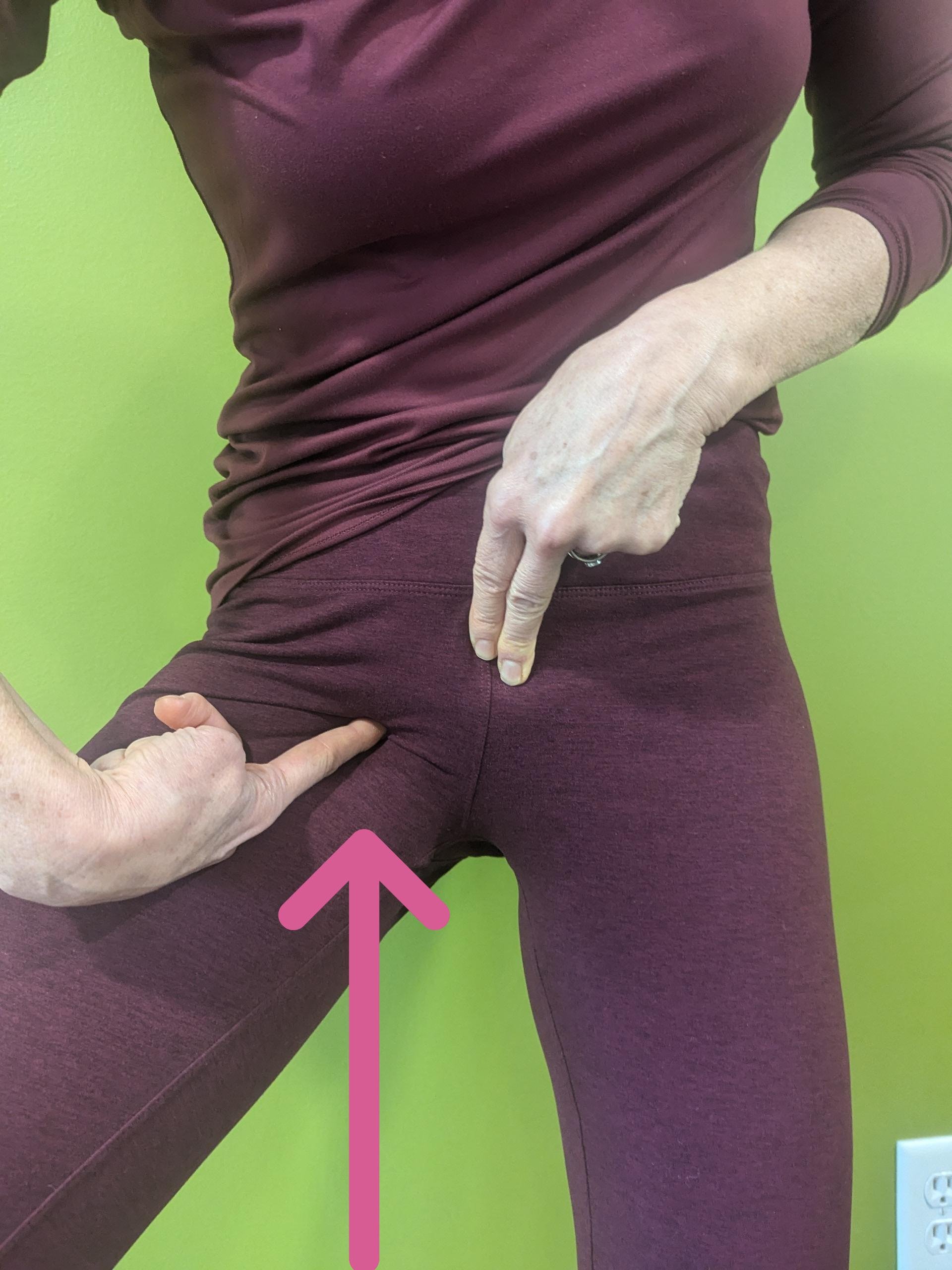
The obturator externus, later referred to as OE is a muscle that is deep in the groin, connecting the anterior inferior part of the pubis to the inside of the upper femur. It's involved in a combination of adduction stability of the leg (and thus can be injured in sharp abduction sprains), with lesser degrees of rotation and flexion. It's not easy to palpate unless you know exactly how to locate certain origin and attachment landmarks, and it connects different parts of our anatomy that make it involved in activities of the leg, trunk and pelvic floor at the same time. The pain pattern usually involves the inside upper thigh, groin, but can also radiate to the perineum and lower abdomen. Mechanism of injury include leg abduction and flexion sprain, slip and trip injuries, weightlifting injuries, and more repetitive type overload injuries such as anterior position of pregnancy and long-distance running.

The reason that the OE is worth talking about is that it can be a source of continued pain even when other parts of an injury have resolved. I have encountered that in many cases where we successfully treated and rehabed sacroiliac and upper thigh injuries for example, but the patient continued to have some very pinpoint groin pain that continue to prevent them from resuming their previous activities. It's often an area that the patients are hesitant to talk about because it will radiate into the genital and pelvic floor area, as well as deep in the inside of the thigh, both areas the patients don't necessarily like to have examined. And an area that many health care providers don’t like to dig in.

Treating and resolving an OE painful syndrome can be a game changer for patient. I'm grateful for my ART training 15 years ago to give me the confidence to properly locate the muscle and perform the appropriate myofascial release on it. The OE responds surprisingly quickly to therapy, usually clearing up within 2 or 3 treatments. It's a little tricky to stretch at home unless you know how to combine the 3 motions to isolate it. I have found my best results with the modified unilateral elevated cobbler with some butterfly flexion.

WHAT ARE THE SCALENE MUSCLES AND WHY DO THEY MATTER ?

It's a question that came up a couple of times this week, always an omen that it's time to blog about it.

The scalenes are a group of 3 muscles, small, but mighty. They are located in the front of the neck, attaching to the anterior lateral aspect of the vertebra and traveling downwards, with one group attaching all the way to the 1st rib just behind and below the clavicle. They are involved in fine motion of the cervical spine such as rotation and lateral bending.

The scalenes are important in many regards:

– as 1 of the primary middle layer of the anterior neck muscles, they are easily injured with rapid extension injuries to the neck which can happen during motor vehicle accidents, falling backwards, and sports. Without involving any additional neurovascular compression, which I'm going to touch on below, the scalenes stem cells can be the source of significant chronic posttraumatic pain, anterior neck posture, and vague radiating pain in the throat and anterior chest

– probably 1 of the most significant contribution of the scalene muscle group is the ability to cause compression over the neurovascular bundle, which is the combination of nerves and arteries and veins that travel between the anterior and middle scalene, just above and then behind the 1st rib, into the arm. This will often result in a sensation of vague pain, tingling, prickly achy sensation in the upper extremity, which does not seem to follow a single nerve root pattern from the cervical spine because the brachial plexus bundle of nerve is comprised of several cervical nerve roots.

https://www.bwclinic.com/blog/2024/11/21/what-are-the-scalene-muscles-and-why-do-they-matter-

CHIROPRACTIC ADJUSTMENT, COORDINATION AND DEXTERITY

I have recently renewed my enthusiasm for digging into chiropractic basic sciences research, especially in the area of neurological efficiency, as my husband discovered the index to chiropractic literature as part of his fellowship in integrative medicine through the Weil Institute in Arizona.

There have been a series of research conducted using very sophisticated tools to measure the efficiency of intracranial signaling speed in patients receiving spinal manipulation. I am extremely grateful for a pioneer Canadian researcher who spent quite a bit of her research career in New Zealand and inspired a whole generation of younger researchers to expand on her earlier work using transcranial magnetic stimulation. To boil it down, the technology has allowed the use of an external magnetic device applied to the skull to modulate speed of transmission between incoming sensory output and outgoing muscular signaling. The technology also allows the researcher to very accurately measure the speed of transmission and the brain. As a general rule, faster transmission results in much more efficient human motor activities such as decreased response time (think athletic performance but also daily activities such as slamming on the brakes to avoid an oncoming vehicle), and overall improved speed and dexterity. This particular study was quite interesting in that it looked at how neck pain affected the speed at which an affected patient could type a series of letters (patients with neck pain were much lower than their counterparts without neck pain), and how a chiropractic adjustment not only restored typing speed to normal in patients with neck pain, but those adjusted individual actually outperformed the control group who had no neck pain and no adjustment.

We often talk about chiropractic for pain alone, but it's important to realize that pain is only a small fraction of our overall neurological functioning. Our earlier chiropractor pioneers were much more interested in the overall neurological function of their patients than pain alone. The study would certainly have reinforced their pursuits, reminding us that chiropractic can be a powerful tool to help humans perform their best, including much more efficient speed dexterity and coordination of the upper extremities for important modern human tasks such as typing.

https://www.researchgate.net/publication/323463302_Subclinical_recurrent_neck_pain_and_its_treatment_impacts_motor_training-induced_plasticity_of_the_cerebellum_and_motor_cortex

SCROLLING TENDINITIS (DE QUERVAIN'S TENOSYNOVITIS)

https://www.youtube.com/shorts/GCtCfrTNej8

I have seen several of these cases recently which then again prompts me to get behind the camera and record a short educational video. Wrist tendinitis can be caused by multiple types of activities, but the increasing number of repetitive strain injuries associated with the use of smart phones, especially single hand phone handling that relies on thumb scrolling, seems to be an increasing percentage. The thumb extensor tendon loops underneath a dorsal wrist ligament called the retinaculum, and the friction of the tendon underneath the retinaculum creates pretty significant inflammation and scarring over time, leading to pinpoint pain on the inside of the wrist during activities that involve wrist extension, holding and picking up objects, and rapidly moving your hand towards the outside of your wrist. When it's becoming an entrenched problem, we have to treat it obviously, but the proverbial ounce of prevention goes a long way in this case. Making sure you scroll with the opposite hand of your phone holding hand will virtually eliminate a lot of left-sided wrist tendinitis, which means that occasionally you have to put on your cup of coffee to use your right index to scroll. Better yet, get off your smart phone and start doing something more beneficial for yourself.

GLP-1 semiglutide medications, muscle and bone loss

Like any clinician working in 21st century American healthcare, over the last 12 months, I've come in contact with an explosion of patients taking GLP-1 semiglutide medications. While those medications were initially labeled for diabetic control, the vast majority of cases presenting in my office are for weight loss. And like many colleagues in the integrated space, balancing the facts about the benefits of a medication addressing severe persistent medically dangerous obesity with known and unknown side effects has been a fine balancing act.

Behind closed doors, when colleagues and I candidly discuss our experience with patients taking this new class of medication, we all wonder when the proverbial other shoe will fall off. There has been an unbridled enthusiasm about the potential for those medication to help curb the scourge of chronic population obesity, but probably a bit of a vacuum when it comes to discussing all of the pros and cons of the prolonged use of a class of medication that historically had been reserved for the patient with poorly controlled diabetes. After 30 years in practice, you acquire a little bit of healthy cynicism when it comes to new treatments of any sort, knowing that some downfalls are not readily available during the "honeymoon" period of a launch that can take months to years to unfold.

There may be a bit of the proverbial shoe starting to drop based on the most recent article published in Lancet (1 of the top dogs of straightforward published medical research). The speed at which patients can lose weight, it turns out, is not all about tissue that you would want to shed. A whopping 3rd of the weight loss comes from lean muscle tissue. This is in contrast to nonmedically induced weight loss from calorie restriction and physical activity, which has a much better track record at preserving lean muscle mass. The latter is quite important for long-term weight maintenance, since muscle tissue is much more metabolically active at burning calories at rest, acting as a blood sugar buffer, among other things.

From a neuromusculoskeletal provider standpoint, the research article hit a bit of a raw nerve because of the implications for our treatment plan. I had already started to suspect that patients on long-term GLP-1 exhibiting significant weight loss were also presenting with decreased overall muscle tone, bulk and endurance, complicating the stabilization phase of the typical spinal and extremity pain that were trying to treat. Another aspect of the rapid weight loss involving muscle tissue that is not brought up by Lancet but is a big concern to me is the concurrent potential impact on decreased bone mass. Lean muscle mass loss and bone loss tend to go hand in hand during significant weight loss.

The research article has reminded me of the importance of reviewing with patients 3 important mitigating strategies with patients on long-term GLP-1:

incorporate a sufficient amount of protein in the diet while on the medication, defined as a minimum of half a gram of protein per pound of body weight. This means that a 200 pound adult would need 100 g of protein. You have to be really intentional about reaching that goal every day through combination of protein dense foods with every meal, and potentially supplemental protein.

Engage in resistance training to 3 times a week that is sufficient to stimulate muscle growth. Just doing activities of daily living and walking is not going to preserve muscle mass.

Consider adding a good bone building support supplement, which is going to contain a combination of bioavailable calcium, magnesium, other bone building minerals, vitamin D3 and vitamin K 2.

3 technique pointers for a safer deadlift

3 TIPS FOR A SAFER DEADLIFT

I've found myself teaching those instructions to patients often enough that I decided to make a quick video about it so I can refer patients to view them again after the appointment. While I have not spent as much time as Dr. Steve in a traditional gym setting, I've done enough deadlifts in my own exercise time to know how quickly they can go wrong, especially if you happen to be recovering from a recent lumbar strain or any other lumbar injury. The main goal of the debt lift modification is to keep a stable flatback in maximum descent and muscular contraction. The 3 modifications, namely wide stance, slightly flexed knees, and firing up the good muscles ahead of time, facilitate maintaining a safe lumbar posture

(photo courtesy of Freepik)

https://www.youtube.com/watch?v=sFqTF5iy3WQ

What causes groin pain ?

It's a question that comes up periodically and since there is no easy answer that I can give to patient during a routine appointment slot, I decided to put my thoughts in writing so I can refer patients to it as a conversation starter.

The groin is a small piece of the body's real estate, that we tend to think of as private, but that can really control your life if it starts acting up.

It's a body area that is the intersection of several structures that can be pain producing, and you need a little bit of attentive detective work to determine the source of the problem.

The main structures that can be involved in producing groin pain:

– referred pain from the middle lumbar area, especially L2 and L3 segments. These would be things like a lumbar disc herniation causing pain to radiate along these dermatomes, or significant bony spurs. The lower lumbar segments such as L4 5 and L5 S1 definitely take the lines shares in terms of referred this pain, so that sometimes we tend to forget that mid lumbar areas can also be a source of referred pain. Patients with groin pain referred from these mid lumbar segments tend to have pain that is aggravated by lumbar movement.

– Referred pain from myofascial structures in the deep hip flexors, especially if involving some peripheral compression of the anterior peripheral nerves that exit that those levels: the femoral nerve, the ilio inguinal nerve, and the lateral femoral continues nerve. This pain can be really tricky to assess, since it will not readily show up on advanced imaging like MRI. During physical examination, deep lateral palpation of those structures can usually be pretty revealing, and the fact that very often activities involving hip flexion can be very triggering.

– The hip joint, or more precisely the acetabular femoral joint (AF joint) . This is the ball and socket joint between the socket in the pelvis (acetabulum) , and the femoral head. This is not usually a joint that is subject to primary misalignments due to its ball and socket nature, although it certainly can, but is subject to articular cartilage degeneration, and tearing and catching of the cartilage rim also known as the labrum. There are specific orthopedic testing that can help isolate the AF joint is the source of the problem, and in addition pain from imaging as well as advanced imaging like MRI can be very diagnostic of the problem starting in that structure. From a history standpoint, patient will often report the sensation of catching or clicking, and pain triggered by hip movement rather than lumbar movement.

– The sacroiliac joint. The sacroiliac is a rather large vertical/horizontal articulation at the base of the sacrum, and the anterior portion of the sacroiliac joint can refer pain to the groin. This would happen with misalignment of the sacroiliac joint internally, that will be often associated with deep pain in the buttocks, difficulty with flexibility in the affected side of the pelvis, and standing with a toe out position on the affected side.

So groin pain can come from many different sources, but it should not be a mystery and a good physical examination can usually point the patient and the treating doctor into the right direction.

Autism spectrum disorders and altered gut microbiome: new scientific diagnostic methods

https://www.nature.com/articles/s41564-024-01739-1?utm_source=klaviyo&utm_medium=email&utm_campaign=%28Email%20-%20Chris%20Kresser%20General%20News%29%20Chris%27s%20Friday%20Favorites&utm_term=new%20study&utm_content=new%20study&_kx=ZpXBDTeEF9QJhwDqQXXrImrT_HpFsBz1ZlYMbsx_Vq0.my75y6

With children going back to school this week, I was reminded of this article I first read earlier in the summer. Children with autism spectrum disorder (ASD) makeup and increasing percentage of our school population, with some debate about the real increasing incidence versus better diagnosis inflating the ranks.

Equally the subject of vigorous debate, is the cause of autism. Or more accurately, the causes, since it appears to be a multifactorial trigger that may be different in various affected individual.

This particular article focused on assessing the microbiome of neuro- typical children versus children with ASD. The integrative health community has long argued that neuro- divergent children almost always will exhibit significant functional G.I. markers abnormalities, which are likely the source of some retrograde brain functioning alteration. That same community has also argued that neuro- divergent children can see improvement in their brain functioning if we can improve their microbiota. This particular research article did not address that question, but clearly confirmed with advanced diagnostic techniques that is the children do indeed have some substantially and statistically significant differences in their digestive ecology. However it's pretty safe to operate on the hypothesis that G.I. intervention that will improve microbiota composition will indeed have a secondary positive effect on the function of the central nervous system. It's especially exciting for chiropractors in the integrative health community, since chiropractic adjustments alone have been shown to improve gut -brain axis signaling, further enhancing the effectiveness of any nutritional intervention.